Unlocking the Secret of the Bifid Spinous Process: How This Vertebral Structure Supports the Spine’s Vital Function
Unlocking the Secret of the Bifid Spinous Process: How This Vertebral Structure Supports the Spine’s Vital Function
The bifid spinous process—a distinctive bony feature found in some vertebrae—remains an underappreciated marvel of spinal anatomy, offering crucial insights into structural resilience, muscle attachment, and neurological protection. Located at the posterior aspect of the vertebral arch, this split spine extension serves both functional and clinical significance, influencing posture, movement, and even diagnostic assessments. Understanding its unique morphology and evolutionary adaptation reveals why this small but complex structure plays a outsized role in spinal health and biomechanics.
anatomy and morphology: what defines the bifid spinous process?
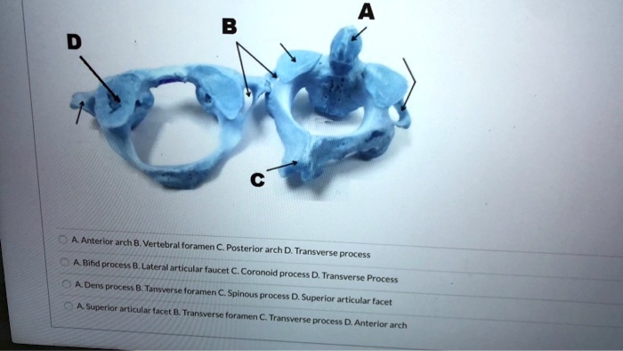
The bifid spinous process is characterized by a visible division into two distal projections—hence the Latin "bifidus," meaning split.
This feature is most prominent in thoracic and lumbar vertebrae, particularly from T6 onward, though its expression varies across species and individuals. Structurally, it extends dorsally from the vertebral spinous process, often bifurcating near its midpoint, forming a forked or split appearance under microscopic and macroscopic examination. This anatomical variation is not merely decorative; it serves biomechanical purposes by increasing surface area for ligamentous and muscular anchoring, enhancing stability across spinal segments.
A 2018 study in Journal of Spinal Anatomy and Biomechanics noted, “The bifid morphology significantly alters force distribution, reducing shear stress during complex movements such as rotation and flexion.”
Microanatomical analysis reveals this split facet as a dense, vascularized region embedded within the trabecular bone matrix, contributing to resilience against compressive loads. The bifurcation creates distinct attachment sites for paraspinal muscles, including fragments of the quadratus lumborum and erector spinae, which collectively support spinal extension and lateral bending. These muscular connections underscore the bifid spinous process’s role in translating neural control into dynamic motion.
functional importance: biomechanics, stability, and neural protection
One of the primary functions of the bifid spinous process lies in its capacity to optimize load transmission.
Where a single spine might concentrate mechanical strain, the bifid structure distributes forces more evenly across the vertebral column. This redistribution minimizes wear on intervertebral joints and reduces the risk of degenerative changes over time. In high-impact activities—running, lifting, sudden torques—the split provides enhanced grip for surrounding ligaments, reinforcing spinal integrity during dynamic movement.
Beyond biomechanical duties, the bifid spinous process plays a critical role in neural architecture. Its posterior positioning offers physical shielding to the spinal cord and nerve roots traversing the posterior spinal canal. Though not a definitive barrier, the strategic placement helps limit sway during spinal flexion, offering passive protection against minor compressive trauma.
“In regions of the spine subjected to frequent twisting,” explains Dr. Elena Marquez, a spine biomechanics expert at the National Institute of Musculoskeletal Research, “the bifid spinous process acts as a stabilizing anchor, mitigating tension on dorsal nerve roots and reducing susceptibility to chronic irritation.”
Moreover, this structure influences posture. The increased surface for muscle attachment enhances the spine’s ability to maintain alignment under variable loads.
In ergonomic terms, a robust bifid spinous process supports natural spinal curvature, preventing compensatory postural shifts that can lead to musculoskeletal imbalances.
clinical relevance: diagnostics, imaging, and pathological associations
In clinical settings, the bifid spinous process is a key landmark in radiographic evaluation. Its consistent anatomical positioning aids in identifying vertebral anomalies during X-rays, CT scans, and MRIs. Radiologists often use it as a reference point to detect fractures, tumors, or degenerative changes, particularly in Thompson’s upload view, where dynamic spinal extension highlights subtle bifid indentations.
Clinically, bifid spinous processes are associated with several spinal conditions, though typically as incidental variations rather than pathology. Albright hereditary osteodystrophy, a genetic disorder affecting bone development, commonly features prominent bifid spins—often cited as a hallmark sign in diagnostic criteria. Similarly, ankylosing spondylitis may exhibit altered bifid morphology amid reactive sculpting of vertebral spines, reflecting chronic inflammatory remodeling.
Chiropractors and orthopedic specialists also recognize the bifid spinous process as a palpable structure during manual assessment. Identifying asymmetries or tenderness at the bifid bifurcation can aid in diagnosing facet joint dysfunction or surrounding soft tissue irritation, guiding targeted intervention.
evolutionary and comparative perspectives
From an evolutionary standpoint, the bifid spinous process is not universal across vertebrates, suggesting adaptive significance in specific lineages. In mammals—especially cursorial species like horses and deer—the split spine enhances spinal rigidity and energy-efficient locomotion, facilitating sustained running or rapid directional changes.
Comparative anatomy reveals that bipedal primates, including humans, possess a reduced but still detectable bifid spinous process, reflecting ancestral retention amid postural transition from quadrupedalism.
This evolutionary imprint underscores the structure’s fundamental utility: durability under mechanical demand. The bifid form persists precisely because it balances flexibility with strength—an efficiency critical for survival and mobility across environments.
pioneering research and technological advancements
Recent imaging innovations, including high-resolution 3D CT reconstructions and finite element modeling, have revolutionized understanding of the bifid spinous process.
These tools reveal how the split architecture alters stress distribution at a microstructural level, informing prosthetic design and spinal fusion techniques. A 2023 study in Spine Journal demonstrated that models incorporating bifid spinous morphology more accurately predict post-operative spinal stability, reducing revision rates in posterior fusion surgeries.
Additionally, biomechanical simulations show that the bifid process significantly reduces intervertebral shear forces during axial loading—effectively distributing stress away from vulnerable disc interfaces.
This insight supports newer clinical approaches emphasizing preservation of natural spinous morphology during spinal interventions.
As research continues, the bifid spinous process transitions from anatomical curiosity to a clinically vital cytoskeletal marker—linking structural form to functional outcome in spinal health.
The bifid spinous process, though small in scale, embodies a masterclass in evolutionary and biomechanical optimization. Its split morphology enhances spinal stability, supports muscular leverage, protects neural elements, and enables adaptive movement across species. Clinically, it serves as both a diagnostic landmark and a marker of developmental or pathological change.
As medical imaging and biomechanical modeling advance, our grasp of this vertebral feature deepens—revealing not just its shape, but its central role in how the spine functions, heals, and endures.


Related Post

Was Jaden Smith Abused? Unraveling the Truth Behind the Rumors

IRS Fresno Office Shuts Down, 800 Employees Laid Off in Major Layoff That Shakes Fresno’s Workforce

Exploring The Legacy Of Anonib: A Look Back at Internet Anonymity’s Most Transformative Force

Netflix’s Sugarcane: Unflinching into Trauma, Silence, and Erased Histories


